Imaging, flow, and samples for discovery research
The exploratory end of pharma R&D moves like an academic bench, not like manufacturing. Conspecta keeps the assay, the images, the flow panels, and the cell lines behind them in one workspace, from the plate to the decision deck.
Built for the exploratory bench
Imaging
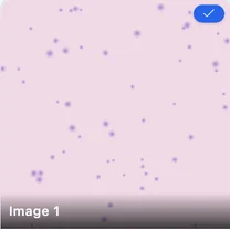
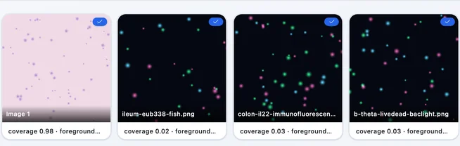
Cell detection, area, intensity, and colocalization on LIF, TIFF, and PNG in the browser, z-stacks included, saved as a reusable analysis attached to the samples it measured. The number in the deck opens back into the analysis that produced it, whoever ran it and wherever they are now.
Flow
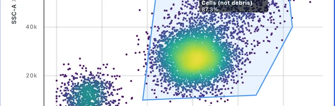
Gating, compensation, and clustering in any browser, with the gating strategy saved as a template the whole group works from. The scientist who needs the answer runs it themselves, the day they need it.
Samples
Samples, cell lines, and treatment conditions carry structured metadata, and every image and data file links back to the material it was measured from. The context sits on the record, where the rest of the program can read it.
Figures
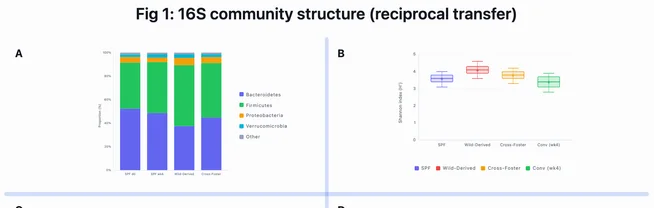
Charts and multi-panel figures are built from the analysis itself rather than pasted beside it, ready for the decision deck and the paper. A curve in the deck traces back to the images behind it, so re-analysis moves the figure with it.
%22%2F%3E%3Cellipse%20cx%3D%22155.4%22%20cy%3D%22135.2%22%20rx%3D%2211.8%22%20ry%3D%227.7%22%20fill%3D%22url(%23n)%22%2F%3E%3Cellipse%20cx%3D%22325.9%22%20cy%3D%22410%22%20rx%3D%228%22%20ry%3D%227.4%22%20fill%3D%22url(%23n)%22%2F%3E%3Cellipse%20cx%3D%22864.8%22%20cy%3D%22557%22%20rx%3D%2210.6%22%20ry%3D%2214.6%22%20fill%3D%22url(%23n)%22%2F%3E%3Cellipse%20cx%3D%22593%22%20cy%3D%22166.5%22%20rx%3D%229%22%20ry%3D%227.5%22%20fill%3D%22url(%23n)%22%2F%3E%3Cellipse%20cx%3D%22866.8%22%20cy%3D%22688.5%22%20rx%3D%2216.2%22%20ry%3D%2210.4%22%20fill%3D%22url(%23n)%22%2F%3E%3Cellipse%20cx%3D%22864.7%22%20cy%3D%22112.4%22%20rx%3D%229.7%22%20ry%3D%2212.6%22%20fill%3D%22url(%23n)%22%2F%3E%3Cellipse%20cx%3D%22108%22%20cy%3D%22564.4%22%20rx%3D%2216.8%22%20ry%3D%2213%22%20fill%3D%22url(%23n)%22%2F%3E%3Cellipse%20cx%3D%22443.7%22%20cy%3D%22658%22%20rx%3D%228.7%22%20ry%3D%228.6%22%20fill%3D%22url(%23n)%22%2F%3E%3Cellipse%20cx%3D%22982.4%22%20cy%3D%22500.3%22%20rx%3D%2211.1%22%20ry%3D%229.5%22%20fill%3D%22url(%23n)%22%2F%3E%3Cellipse%20cx%3D%22281.9%22%20cy%3D%22714.3%22%20rx%3D%2215.1%22%20ry%3D%2211.6%22%20fill%3D%22url(%23n)%22%2F%3E%3Cellipse%20cx%3D%22617.2%22%20cy%3D%22695.3%22%20rx%3D%226.3%22%20ry%3D%229.6%22%20fill%3D%22url(%23n)%22%2F%3E%3Cellipse%20cx%3D%221238%22%20cy%3D%22545.8%22%20rx%3D%227.4%22%20ry%3D%228.4%22%20fill%3D%22url(%23n)%22%2F%3E%3Cellipse%20cx%3D%22325.6%22%20cy%3D%22209.7%22%20rx%3D%226.9%22%20ry%3D%2211.7%22%20fill%3D%22url(%23n)%22%2F%3E%3Cellipse%20cx%3D%221210.7%22%20cy%3D%22202.4%22%20rx%3D%2210.3%22%20ry%3D%228.3%22%20fill%3D%22url(%23n)%22%2F%3E%3Cellipse%20cx%3D%221204.8%22%20cy%3D%22307.9%22%20rx%3D%2211.8%22%20ry%3D%2216.4%22%20fill%3D%22url(%23n)%22%2F%3E%3Cellipse%20cx%3D%22711.5%22%20cy%3D%22654.2%22%20rx%3D%226.5%22%20ry%3D%227.6%22%20fill%3D%22url(%23n)%22%2F%3E%3Cellipse%20cx%3D%221041.1%22%20cy%3D%22647.8%22%20rx%3D%2211.4%22%20ry%3D%229.4%22%20fill%3D%22url(%23n)%22%2F%3E%3Cellipse%20cx%3D%221113.7%22%20cy%3D%22101.1%22%20rx%3D%2211.1%22%20ry%3D%2212.6%22%20fill%3D%22url(%23n)%22%2F%3E%3Cellipse%20cx%3D%22388.6%22%20cy%3D%22449.3%22%20rx%3D%2210.9%22%20ry%3D%227.9%22%20fill%3D%22url(%23n)%22%2F%3E%3Cellipse%20cx%3D%22538.6%22%20cy%3D%22309.8%22%20rx%3D%2213%22%20ry%3D%228.6%22%20fill%3D%22url(%23n)%22%2F%3E%3Cellipse%20cx%3D%22863.5%22%20cy%3D%22308.7%22%20rx%3D%2218.2%22%20ry%3D%2212.2%22%20fill%3D%22url(%23n)%22%2F%3E%3Cellipse%20cx%3D%22393%22%20cy%3D%22366.3%22%20rx%3D%2212.3%22%20ry%3D%2212.1%22%20fill%3D%22url(%23n)%22%2F%3E%3Cellipse%20cx%3D%22308.2%22%20cy%3D%22438.9%22%20rx%3D%2213.3%22%20ry%3D%228.9%22%20fill%3D%22url(%23n)%22%2F%3E%3Cellipse%20cx%3D%22265.1%22%20cy%3D%22179.3%22%20rx%3D%228.7%22%20ry%3D%227.9%22%20fill%3D%22url(%23n)%22%2F%3E%3Cellipse%20cx%3D%2265.9%22%20cy%3D%22718.5%22%20rx%3D%2211.5%22%20ry%3D%228%22%20fill%3D%22url(%23n)%22%2F%3E%3Cellipse%20cx%3D%22801.3%22%20cy%3D%22281.5%22%20rx%3D%2212.1%22%20ry%3D%229.7%22%20fill%3D%22url(%23n)%22%2F%3E%3Cellipse%20cx%3D%22740.1%22%20cy%3D%22392.7%22%20rx%3D%2211.9%22%20ry%3D%228.5%22%20fill%3D%22url(%23n)%22%2F%3E%3Cellipse%20cx%3D%22573.3%22%20cy%3D%22295.8%22%20rx%3D%2210.5%22%20ry%3D%229.4%22%20fill%3D%22url(%23n)%22%2F%3E%3Cellipse%20cx%3D%22755.2%22%20cy%3D%22560.8%22%20rx%3D%2210.6%22%20ry%3D%229.6%22%20fill%3D%22url(%23n)%22%2F%3E%3Cellipse%20cx%3D%22267.8%22%20cy%3D%22627.6%22%20rx%3D%2217.3%22%20ry%3D%2211.6%22%20fill%3D%22url(%23n)%22%2F%3E%3Cellipse%20cx%3D%221130.4%22%20cy%3D%22447.5%22%20rx%3D%2215.2%22%20ry%3D%2210.1%22%20fill%3D%22url(%23n)%22%2F%3E%3Cellipse%20cx%3D%22203.4%22%20cy%3D%22234.4%22%20rx%3D%2210.7%22%20ry%3D%227.7%22%20fill%3D%22url(%23n)%22%2F%3E%3Cellipse%20cx%3D%2275.2%22%20cy%3D%22725.6%22%20rx%3D%229%22%20ry%3D%2212.7%22%20fill%3D%22url(%23n)%22%2F%3E%3Cellipse%20cx%3D%221256.6%22%20cy%3D%22491.8%22%20rx%3D%228.2%22%20ry%3D%2212.8%22%20fill%3D%22url(%23n)%22%2F%3E%3Cellipse%20cx%3D%221218.6%22%20cy%3D%22552.6%22%20rx%3D%229.1%22%20ry%3D%229.9%22%20fill%3D%22url(%23n)%22%2F%3E%3Cellipse%20cx%3D%221211.5%22%20cy%3D%2248.9%22%20rx%3D%2213.7%22%20ry%3D%229.9%22%20fill%3D%22url(%23n)%22%2F%3E%3Cellipse%20cx%3D%22545.5%22%20cy%3D%22570.3%22%20rx%3D%228.7%22%20ry%3D%2212.1%22%20fill%3D%22url(%23n)%22%2F%3E%3Cellipse%20cx%3D%221253.7%22%20cy%3D%22163.9%22%20rx%3D%2212.4%22%20ry%3D%229.3%22%20fill%3D%22url(%23n)%22%2F%3E%3Cellipse%20cx%3D%22381.2%22%20cy%3D%22293.3%22%20rx%3D%226.8%22%20ry%3D%227.3%22%20fill%3D%22url(%23n)%22%2F%3E%3Cellipse%20cx%3D%22478.4%22%20cy%3D%22450.7%22%20rx%3D%228.7%22%20ry%3D%2212.6%22%20fill%3D%22url(%23n)%22%2F%3E%3Cellipse%20cx%3D%22187.2%22%20cy%3D%22493%22%20rx%3D%2210.3%22%20ry%3D%2210.5%22%20fill%3D%22url(%23n)%22%2F%3E%3Cellipse%20cx%3D%22140.8%22%20cy%3D%2279.2%22%20rx%3D%227.5%22%20ry%3D%2212.6%22%20fill%3D%22url(%23n)%22%2F%3E%3Cellipse%20cx%3D%221214.1%22%20cy%3D%2267.1%22%20rx%3D%2211.4%22%20ry%3D%228%22%20fill%3D%22url(%23n)%22%2F%3E%3Cellipse%20cx%3D%22749.8%22%20cy%3D%22731.1%22%20rx%3D%228.8%22%20ry%3D%227.2%22%20fill%3D%22url(%23n)%22%2F%3E%3Cellipse%20cx%3D%22353%22%20cy%3D%22185.4%22%20rx%3D%2211.6%22%20ry%3D%229.8%22%20fill%3D%22url(%23n)%22%2F%3E%3Cellipse%20cx%3D%22787.5%22%20cy%3D%22338.4%22%20rx%3D%227.3%22%20ry%3D%2212.6%22%20fill%3D%22url(%23n)%22%2F%3E%3Cellipse%20cx%3D%22909.2%22%20cy%3D%22357.8%22%20rx%3D%229.6%22%20ry%3D%2213.5%22%20fill%3D%22url(%23n)%22%2F%3E%3Cellipse%20cx%3D%22866%22%20cy%3D%22270.2%22%20rx%3D%229.5%22%20ry%3D%2216%22%20fill%3D%22url(%23n)%22%2F%3E%3Cellipse%20cx%3D%22309.4%22%20cy%3D%22661%22%20rx%3D%229.6%22%20ry%3D%2210.5%22%20fill%3D%22url(%23n)%22%2F%3E%3Cellipse%20cx%3D%221070.5%22%20cy%3D%22632.4%22%20rx%3D%228.3%22%20ry%3D%228.7%22%20fill%3D%22url(%23n)%22%2F%3E%3Cellipse%20cx%3D%22523.5%22%20cy%3D%22142.5%22%20rx%3D%228%22%20ry%3D%2210.6%22%20fill%3D%22url(%23n)%22%2F%3E%3Cellipse%20cx%3D%22460.4%22%20cy%3D%22174.8%22%20rx%3D%2212%22%20ry%3D%2212.5%22%20fill%3D%22url(%23n)%22%2F%3E%3Cellipse%20cx%3D%221114.4%22%20cy%3D%22373%22%20rx%3D%229%22%20ry%3D%2214.5%22%20fill%3D%22url(%23n)%22%2F%3E%3Cellipse%20cx%3D%22504.3%22%20cy%3D%22344.7%22%20rx%3D%228.8%22%20ry%3D%2210%22%20fill%3D%22url(%23n)%22%2F%3E%3Cellipse%20cx%3D%22893.5%22%20cy%3D%22545.6%22%20rx%3D%2210.5%22%20ry%3D%2213.8%22%20fill%3D%22url(%23n)%22%2F%3E%3Cellipse%20cx%3D%22111.8%22%20cy%3D%22262%22%20rx%3D%2211.2%22%20ry%3D%227.7%22%20fill%3D%22url(%23n)%22%2F%3E%3Cellipse%20cx%3D%221021.1%22%20cy%3D%2251.9%22%20rx%3D%2212.5%22%20ry%3D%228.9%22%20fill%3D%22url(%23n)%22%2F%3E%3Cellipse%20cx%3D%221193.6%22%20cy%3D%22351.6%22%20rx%3D%2213.3%22%20ry%3D%2210.2%22%20fill%3D%22url(%23n)%22%2F%3E%3Cellipse%20cx%3D%221107.9%22%20cy%3D%22523.3%22%20rx%3D%2211.7%22%20ry%3D%2210.2%22%20fill%3D%22url(%23n)%22%2F%3E%3Cellipse%20cx%3D%22579.2%22%20cy%3D%22483.3%22%20rx%3D%2211.8%22%20ry%3D%229.5%22%20fill%3D%22url(%23n)%22%2F%3E%3Cellipse%20cx%3D%22273.9%22%20cy%3D%22492.6%22%20rx%3D%2214.2%22%20ry%3D%229.7%22%20fill%3D%22url(%23n)%22%2F%3E%3Cellipse%20cx%3D%22342.2%22%20cy%3D%2241.2%22%20rx%3D%228.6%22%20ry%3D%229.3%22%20fill%3D%22url(%23n)%22%2F%3E%3Cellipse%20cx%3D%221175.7%22%20cy%3D%22177.2%22%20rx%3D%2212.1%22%20ry%3D%228%22%20fill%3D%22url(%23n)%22%2F%3E%3Cellipse%20cx%3D%221009.1%22%20cy%3D%22162.2%22%20rx%3D%2212.8%22%20ry%3D%2213.1%22%20fill%3D%22url(%23n)%22%2F%3E%3Cellipse%20cx%3D%22908%22%20cy%3D%22712%22%20rx%3D%228.3%22%20ry%3D%2213.4%22%20fill%3D%22url(%23n)%22%2F%3E%3Cellipse%20cx%3D%221159.4%22%20cy%3D%22558.7%22%20rx%3D%2210.5%22%20ry%3D%228.9%22%20fill%3D%22url(%23n)%22%2F%3E%3Cellipse%20cx%3D%22154.6%22%20cy%3D%2279.7%22%20rx%3D%228.5%22%20ry%3D%229.6%22%20fill%3D%22url(%23n)%22%2F%3E%3Cellipse%20cx%3D%22224.4%22%20cy%3D%22358.1%22%20rx%3D%2211.6%22%20ry%3D%2212.6%22%20fill%3D%22url(%23n)%22%2F%3E%3Cellipse%20cx%3D%221051.7%22%20cy%3D%22167.9%22%20rx%3D%2211.6%22%20ry%3D%227.5%22%20fill%3D%22url(%23n)%22%2F%3E%3Cellipse%20cx%3D%2272%22%20cy%3D%22469.1%22%20rx%3D%2210.6%22%20ry%3D%227.9%22%20fill%3D%22url(%23n)%22%2F%3E%3Cellipse%20cx%3D%221191.6%22%20cy%3D%22274.6%22%20rx%3D%228.9%22%20ry%3D%229.4%22%20fill%3D%22url(%23n)%22%2F%3E%3Cellipse%20cx%3D%22817.4%22%20cy%3D%22690.6%22%20rx%3D%2211.1%22%20ry%3D%2210.6%22%20fill%3D%22url(%23n)%22%2F%3E%3Cellipse%20cx%3D%2265.6%22%20cy%3D%22571.6%22%20rx%3D%228.8%22%20ry%3D%229.5%22%20fill%3D%22url(%23n)%22%2F%3E%3Cellipse%20cx%3D%22255.4%22%20cy%3D%22307.3%22%20rx%3D%2211.3%22%20ry%3D%2214.1%22%20fill%3D%22url(%23n)%22%2F%3E%3Cellipse%20cx%3D%221235%22%20cy%3D%22149.2%22%20rx%3D%2211.8%22%20ry%3D%228.4%22%20fill%3D%22url(%23n)%22%2F%3E%3Cellipse%20cx%3D%221248.8%22%20cy%3D%2267.6%22%20rx%3D%2212.1%22%20ry%3D%229.9%22%20fill%3D%22url(%23n)%22%2F%3E%3Cellipse%20cx%3D%22846.1%22%20cy%3D%22688.1%22%20rx%3D%228.8%22%20ry%3D%229.3%22%20fill%3D%22url(%23n)%22%2F%3E%3Cellipse%20cx%3D%22546.3%22%20cy%3D%22233.5%22%20rx%3D%2213.7%22%20ry%3D%2211.2%22%20fill%3D%22url(%23n)%22%2F%3E%3Cellipse%20cx%3D%22630.1%22%20cy%3D%22705.8%22%20rx%3D%2212.1%22%20ry%3D%2211.5%22%20fill%3D%22url(%23n)%22%2F%3E%3Cellipse%20cx%3D%22664.5%22%20cy%3D%22343.7%22%20rx%3D%229.4%22%20ry%3D%2210.4%22%20fill%3D%22url(%23n)%22%2F%3E%3Cellipse%20cx%3D%22725.5%22%20cy%3D%22286.4%22%20rx%3D%2212.1%22%20ry%3D%2210.1%22%20fill%3D%22url(%23n)%22%2F%3E%3Cellipse%20cx%3D%22876.3%22%20cy%3D%22290.9%22%20rx%3D%2212.1%22%20ry%3D%2211.7%22%20fill%3D%22url(%23n)%22%2F%3E%3Cellipse%20cx%3D%22738.6%22%20cy%3D%22569.4%22%20rx%3D%2212.6%22%20ry%3D%2211.6%22%20fill%3D%22url(%23n)%22%2F%3E%3Cellipse%20cx%3D%22673.6%22%20cy%3D%22714.2%22%20rx%3D%2213.5%22%20ry%3D%2210.2%22%20fill%3D%22url(%23n)%22%2F%3E%3Cellipse%20cx%3D%22431.3%22%20cy%3D%22521.4%22%20rx%3D%2211.1%22%20ry%3D%228.8%22%20fill%3D%22url(%23n)%22%2F%3E%3Cellipse%20cx%3D%22133.3%22%20cy%3D%22269.9%22%20rx%3D%2210.4%22%20ry%3D%229.5%22%20fill%3D%22url(%23n)%22%2F%3E%3Cellipse%20cx%3D%22264.1%22%20cy%3D%22433.8%22%20rx%3D%2213.1%22%20ry%3D%2211.8%22%20fill%3D%22url(%23n)%22%2F%3E%3Cellipse%20cx%3D%22574.8%22%20cy%3D%22456.5%22%20rx%3D%229.4%22%20ry%3D%2216.1%22%20fill%3D%22url(%23n)%22%2F%3E%3Cellipse%20cx%3D%221071.6%22%20cy%3D%22301.6%22%20rx%3D%221.5%22%20ry%3D%221.1%22%20fill%3D%22url(%23n)%22%2F%3E%3Cellipse%20cx%3D%22459.3%22%20cy%3D%22408.6%22%20rx%3D%224.2%22%20ry%3D%222.7%22%20fill%3D%22url(%23n)%22%2F%3E%3Cellipse%20cx%3D%22840.7%22%20cy%3D%22310.8%22%20rx%3D%221.1%22%20ry%3D%221.9%22%20fill%3D%22url(%23n)%22%2F%3E%3Cellipse%20cx%3D%22312.1%22%20cy%3D%22329.6%22%20rx%3D%222.5%22%20ry%3D%223.4%22%20fill%3D%22url(%23n)%22%2F%3E%3Cellipse%20cx%3D%22972.4%22%20cy%3D%22157.6%22%20rx%3D%222.4%22%20ry%3D%221.6%22%20fill%3D%22url(%23n)%22%2F%3E%3Cellipse%20cx%3D%22975.1%22%20cy%3D%22474.6%22%20rx%3D%223.2%22%20ry%3D%225.2%22%20fill%3D%22url(%23n)%22%2F%3E%3Cellipse%20cx%3D%22792%22%20cy%3D%22697.5%22%20rx%3D%222.5%22%20ry%3D%221.8%22%20fill%3D%22url(%23n)%22%2F%3E%3Cellipse%20cx%3D%221137.9%22%20cy%3D%22656.5%22%20rx%3D%223.1%22%20ry%3D%223.4%22%20fill%3D%22url(%23n)%22%2F%3E%3Cellipse%20cx%3D%22186.5%22%20cy%3D%2241.6%22%20rx%3D%224.7%22%20ry%3D%223.1%22%20fill%3D%22url(%23n)%22%2F%3E%3Cellipse%20cx%3D%22113%22%20cy%3D%22521.8%22%20rx%3D%221.8%22%20ry%3D%222.9%22%20fill%3D%22url(%23n)%22%2F%3E%3Cellipse%20cx%3D%2273.3%22%20cy%3D%22589.1%22%20rx%3D%224.2%22%20ry%3D%223.2%22%20fill%3D%22url(%23n)%22%2F%3E%3Cellipse%20cx%3D%22181.5%22%20cy%3D%22644.9%22%20rx%3D%221.9%22%20ry%3D%223%22%20fill%3D%22url(%23n)%22%2F%3E%3Cellipse%20cx%3D%221183.4%22%20cy%3D%22223.7%22%20rx%3D%221.6%22%20ry%3D%221.4%22%20fill%3D%22url(%23n)%22%2F%3E%3Cellipse%20cx%3D%221252.5%22%20cy%3D%22459.9%22%20rx%3D%223%22%20ry%3D%223.3%22%20fill%3D%22url(%23n)%22%2F%3E%3C%2Fsvg%3E)
Questions pharma teams ask
Is Conspecta GxP compliant, or does it support 21 CFR Part 11?
The standard service is research software, and it isn't validated. Notes, protocols, and sequences in Genetics carry version history. Changes are logged with who made them and when, and the log cannot be edited. Sign-offs record who approved what and show when the record has changed since, and protocol runs keep a step-by-step trail. A Business engagement validates Conspecta against your workflow and adds the signature controls Part 11 requires. Talk to us before a GLP, GMP, or QC release workflow moves in.
Can we put clinical or patient data in Conspecta?
Our standard terms exclude protected health information, so patient data and clinical trial records do not belong in the standard service. Identifiable patient data needs a BAA, which is quoted with a Business plan. Data is stored and processed in the United States, and another region is quoted the same way, so talk to us before anything covered moves.
Can each program be kept separate from the others?
Yes. Each program lives in its own project with its own membership, and projects are isolated at the database level, so people working on one series do not see the rest of the portfolio. Exports run per project too, which matters when a contractor or an academic collaborator needs one slice of the work.
How is Conspecta priced for an industry team?
Commercial teams are quoted per seat, and seats, storage, and the monthly usage allowance are all settled in that quote. Support for regulated workflows is quoted in that same conversation rather than sold self-serve. A scientist can start on the free plan and test it with real data first.
Will our data be locked in?
No. Images come out at full resolution, samples, statistics, and analysis results as Excel, gated populations as FCS files, sequences as GenBank, and figures as PNG or SVG. Exports are free and never count toward your usage, and the full project export carries every sign-off and the provenance links behind a result, so a program that ever moves takes its history with it.
Start with one assay
Bring one assay in and see the readout, the analysis behind it, and the figure it becomes stay connected. No IT project to open.